Medical Engineering Collaboration / Life Sciences
Luminescent material for a highly-sensitive in vivo imaging that can visualize to a depth of 5-6cm from the epidermis
Overview
Organization Name
Maki Shojiro, Associate Professor of Center of Neuroscience and Biomedical Engineering, Graduate School of Informatics and Engineering, Department of Engineering Science, The University of Electro-Communications
Documentation
Summary
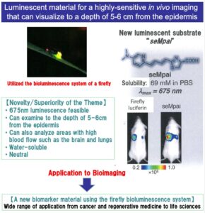
In recent years, “Bioimaging”, which can observe the development process of disease in real-time by using fluorescent agents (bioprobes) to illuminate intracellular proteins, has been attracting attention. In this study, we developed a luminescent substrate that gives an emission peak within the biological window (650 nm ~900 nm) at which “imaging to a depth of 5-6cm from the epidermis is possible”, using the bioluminescent system of a firefly. The substrate is neutral and highly soluble, and can also be used to analyze the areas with high blood flow such as the brain and lungs. It can be expected to be used not only in oncology and regenerative medicine, but also in bioluminescence imaging in organisms such as miniature pigs and marmosets. Any company or laboratory that is willing to utilize this technology is welcome.
Details
Simplified Diagram
Background
“Bioimaging” is known as a technology that illuminates and observes metastatic cancer by illuminating intracellular proteins using fluorescent materials. A labeling material (called a bioprobe) that can detect specific cells by fluorescence or luminescence is essential for such bioimaging technology. However, in vivo, emission less than 650 nm does not penetrate to the outside of the body that in order to precisely carry out imaging of deep layer tissues in a living organism, a material of more than 650 nm is required. Precise imaging of deep layer tissues in living organisms requires materials with the emission above 650 nm.
In this study, we propose a new labeling material “seMpai” that emits light at 675 nm, based on the knowledge obtained from the firefly luminescence system.
Any company or laboratory that is willing to utilize this technology is welcome.
Technical content
There are about 800 types of organisms that emit light such as fireflies and sea-fireflies. Recently, attention has been focused on the fact that these bioluminescent organisms can achieve high energy conversion efficiency. For example, the energy conversion efficiency of the steam engine, which is said to have the highest conversion efficiency among those produced by humans, is 30~40%, while that of fireflies is said to be about 41%. While the bioluminescence of fireflies has excellent luminous efficiency, it has a weak point that the emission color (wavelength) is limited.
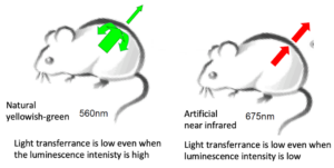
Luciferin, a luminescent substrate derived from fireflies, glows yellow when it chemically reacts with the luminescent enzyme “luciferase.” This reaction has already been widely used in life sciences and other kinds of researches in the world, but it shines in yellowish-green (emission wavelength; 560 nm). Besides fireflies, luminescence materials from bioluminescent organisms such as sea-fireflies and bioluminescent shrimp are also used, but they are still utilized for the same color as in nature (blue), and the long-wavelength (red) luminescent material is still not commercially available.
In the Maki Lab, we have achieved long-wavelength emission (red) of more than 675 nm, using our unique know-how to artificially modify the luminescent substrate (luciferin). Until today, Maki Lab has developed and launched “Akarumine ®” and “TokeOni.” We have already succeeded in light emission imaging in miniature pigs and marmosets using TokeOni.
Strengths of technology and know-how (innovation, superiority, utility)
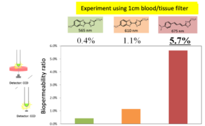
Light emission in the long-wavelength region of 650 nm~900 nm is called the “biological window.” Since emission in this region has high biological permeability (=high light transmission), even emission with very low intensity or from deep tissue layer can be easily visualized from the outside.
*High biopermeability at 675nm.
By using an emission wavelength with high permeability to biological tissue, highly-sensitive imaging can be realized and “imaging to a depth of 5-6cm from the epidermis is possible”. This makes it feasible to analyze areas with high blood flow such as the brain and lungs.
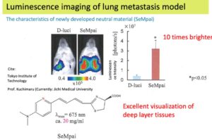
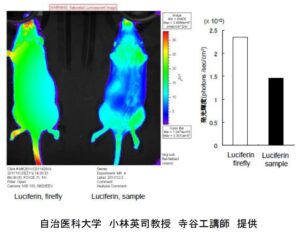
The results of luminescence imaging of a lung metastasis model using the newly developed and launched labeling material (SeMpai) are as below. The advantage of long-wavelength emission is effective, and even the lungs, which are deep layer tissues, can be visualized.
As it can be administered as neutral, the blood does not become acidic. Due to its high solubility, concentration adjustment is easy that high-concentration administration is possible.
Image of Allied Company
We welcome companies that are willing to use this technology.
For example, we can partner with the following companies.
1) Companies / laboratories that want to perform in vivo imaging on medium-sized animals such as miniature pigs, common marmoset, and monkeys.
2) Companies / laboratories that want to image deep tissue areas, such as the brain and lungs.
3) Pharmaceutical manufacturers / laboratories wishing to utilize long-wavelength luminescent bioprobes.
4) Other companies with technical issues related to bioluminescence.
Utilization of technology and know-how (image)
The material can be used as a bioprobe in a wide range of applications such as tumor applications and regenerative medicine applications (application to IPS cells). For example, the following procedure can be used to analyze the transfer phenomenon of oncogenes.
1) Incorporate the firefly luciferase gene (firefly luciferase) into “cultured cancer cells”, and then transplant into experimental animals such as mice to artificially produce cancer.
2) When the long-wavelength bioprobe is administered from the outside of the body, the chemical reaction between the enzyme and the substrate occurs only in the cancer cells, and long-wavelength luminescence (red) is generated.
3) Observe the emission pattern with an imaging device.
*The red light of the long-wavelength is invisible to the human eye, but can be observed with an experimental measurement device. In addition, since the observation with the living organism can be continued for many days, it is possible to track how cancer develops, grows, and metastasizes at a cellular level.
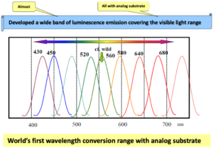
Also, the artificial firefly luminescence system can be used to not only make biomarkers with long-wavelength light emission but also to emit a band of wavelength (=multi-color) with a wide range over RGB by customizing the substrate.
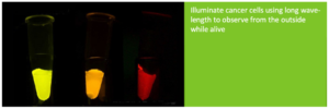
*Multi-color emission using the artificial firefly luminescence system.
The red (right) can be used as a bioprobe.
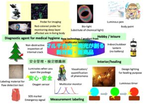
*Applications of multi-color emission
Flow of Technology and Know-How Application
After your inquiry, we will explain detailed technical contents at the technical meeting. We can also provide various technical consultations on bioluminescence and organic synthesis, not only limited to this study.
Description of the Technical Terms
Bioprobe
Also called labeling material, it refers to low molecular weight organic compounds that are useful for analyzing biological functions. The bioprobe enables to obtain useful insights into life sciences, such as visualization of gene expression in cells.
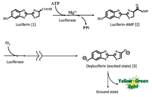
The principle of firefly bioluminescence
Bioluminescence of fireflies is produced when luminescent enzyme (luciferase) acts on a luminescent substrate (luciferin) and undergoes an oxidation reaction using oxygen in the air.
Due to the high energy conversion efficiency of bioluminescence, researches are being carried out around the world, but most of them are on luciferase.
Doctor Maki has know-how and novelty for the artificial synthesis of the luminescent substrate itself.
The bioluminescence of fireflies is carried out through a two-step process.

