Simplified
Background
In the medical field, it is pointed out that the shortage of the pathologist has become chronic, and that the patient may take a long time to obtain the diagnosis results, and that the erroneous diagnosis may increase due to excessive over diagnosis of the pathologist. Therefore, in recent years, a tool to assist in pathological diagnosis has been developed using AI. However, the detection accuracy is not sufficient yet.
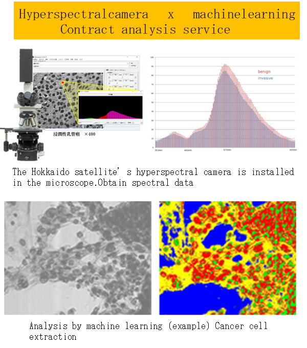
The hyperspectral camera developed by Hokkaido Satellite can obtain nearly 40 times more data than normal digital images, recent improvements in computational power of CPUs have allowed us to analyze the large-scale data of this hyper spectrum without artificially reducing the large scale data of the spectrum. Moreover, the use of general-purpose programming language, which is suitable for the use of machine learning, such as Python and R, has led to the development of a system that combines the conventional digital images with the hyper spectrum and the original AI, and has been applied to the pathological diagnosis of the system.
Technical Content
Technology overview
The data captured by a hyperspectral camera is the spectral information of the usual digital image RGB 3 band divided into 141 bands, which is nearly 40 times more than the normal digital image.
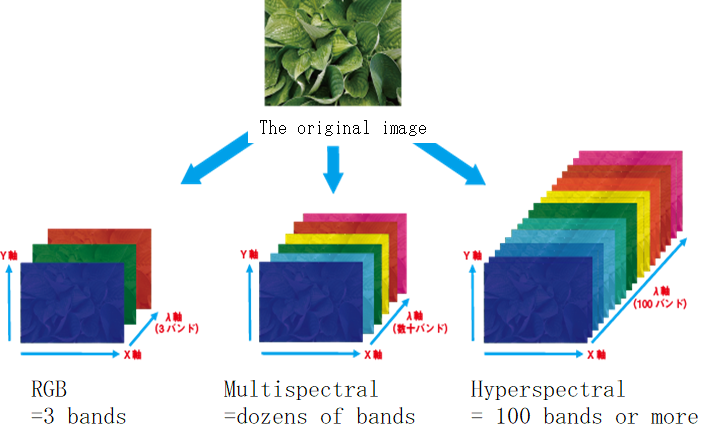
By using the machine learning developed by the Hokkaido satellite in the hyperspectral image, it is possible to find minute structural changes and defects.
The “ANSWER,” a combination of this hyper spectrum and its unique AI, can be analyzed by machine learning only with several hyperspectral images, and can be classified not only by cancer, non-cancer classification, but also by sub-classification.
| ANSWER | Other company’s product | |
| Data | Hyper spectrum | RGB image |
| Analysis | AI (original method) | AI (deep running) |
| Learning data | Data volume: 1 | Data amount: 100 |
| Accuracy | Sub-classification is also possible | It can be classified as cancer or non-cancer |
The developed AI algorithm provides a clear basis for the test results (patented). At the time of pathological diagnosis, we got almost 99% of identification rate for pancreatic cancer, breast cancer, ovarian cancer, prostate cancer, colon cancer, and so on.
Analysis Example
- Identify Malignant Tumors
Malignant tumors are eventually diagnosed as “cancer” by microscopic observation of a pathologist. In particular, tumor of the pancreas is difficult to identify, and objective diagnostic index is required. By measuring microscopically magnified pancreatic cells with a hyperspectral camera, we succeeded in obtaining an accuracy of 98.7% at the cellular level. The analysis technique uses a machine learning technique such as SVM and clustering.
Example of Pancreatic Ductal Carcinoma
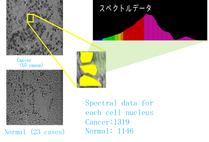
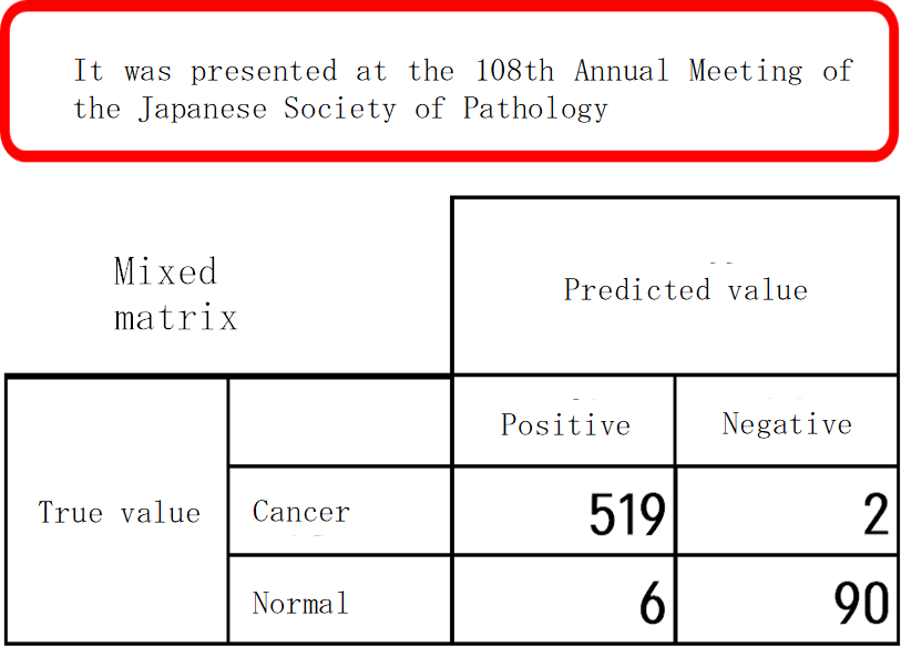
Results : Answer rate 98.7%, sensitivity 97.8%, specificity 98.9%
- High precision detection of nuclei

We can count and extract nuclear nuclei. Compared to normal digital images,
We can identify the boundary of the nucleus accurately.
Example of predictive analysis by dedicated application
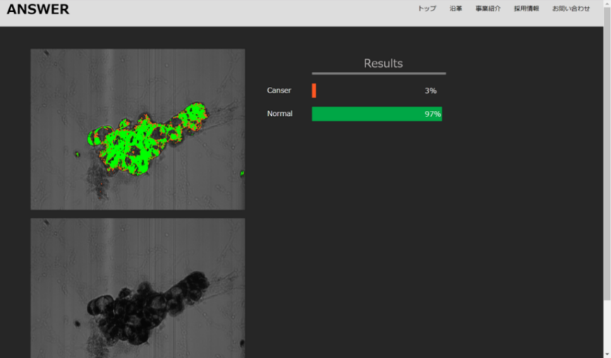
Flow of analysis by dedicated application
It’s easy to analyze by dragging and dropping HSI data on Web apps.
(Required time 1 minute).
- Measurement of hyperspectral images (HSI)
- Upload HSI to “ANSWER”
- “ANSWER” indicates a place where a cancer is likely to be a cancer, and a pathologist’s final judgment.
【Analysis service】
You will send the sample you would like to analyze, and we send back a report on the results. It is available for both stained sample and non-stained sample. In case of analyzing them through a microscope, All samples should be on glass slides.
Strengths of technologies and know-how (novelty, superiority, utility)
We can obtain high accuracy with fewer samples. Hyperspectral data has 40 times more information than normal digital images. By increasing the amount of information available from the one sample, it is possible to obtain high precision in the sample.
Image of a cooperative company
1) Companies and laboratories that are researching and testing image processing by image processing
2) Hospitals and laboratories for pathological diagnosis
3) Companies that want to detect foreign matter and quantify quality by hyper-spectrum
Utilization of technologies and know-how (images)
■Examples of pathological diagnosis
It is possible to screen cancer cells mixed with the cells removed from the patient. When a sample with a high likelihood of cancer is diagnosed preferentially by the pathologist, it is possible to reduce the burden on the business and improve the accuracy of the diagnosis.
■Various scenes that used a hyperspectral camera
AI can also learn and analyze foreign substances in food, quantify components, classify freshness, etc.
Flow of technology and know-how
If you are interested in the use of this technology, please feel free to contact us.
Description of the technical terms
[ANSWER]
Abbreviation for Analyze Nature & Space with Electromagnet Radiation.
[SVM]
Abbreviation for Support Vector Machine.
A method of identifying recent neighbor sample plots from the identification plane by optimizing the identification plane as a support vector.
[Clustering]
It is an algorithm that defines a distance function of a cluster called a cluster by defining a distance function.
[Sub-Classification]
The pathological classification is largely classified into malignant grade and histological type.
Furthermore, various classifications are sub-classification.
Pancreatic carcinoma
It is a malignant tumor that develops in the pancreatic duct in pancreatic cancer.

